A mole that has changed in color, shape, size, or sensation usually deserves more than a quick glance. When a dermatologist recommends a mole biopsy procedure, the goal is straightforward – get a clear diagnosis and decide whether any further treatment is needed.
For many patients, the hardest part is not the procedure itself. It is the uncertainty beforehand. Knowing what the visit involves, why one biopsy method is chosen over another, and what happens after the sample is sent to the lab can make the process feel much more manageable.
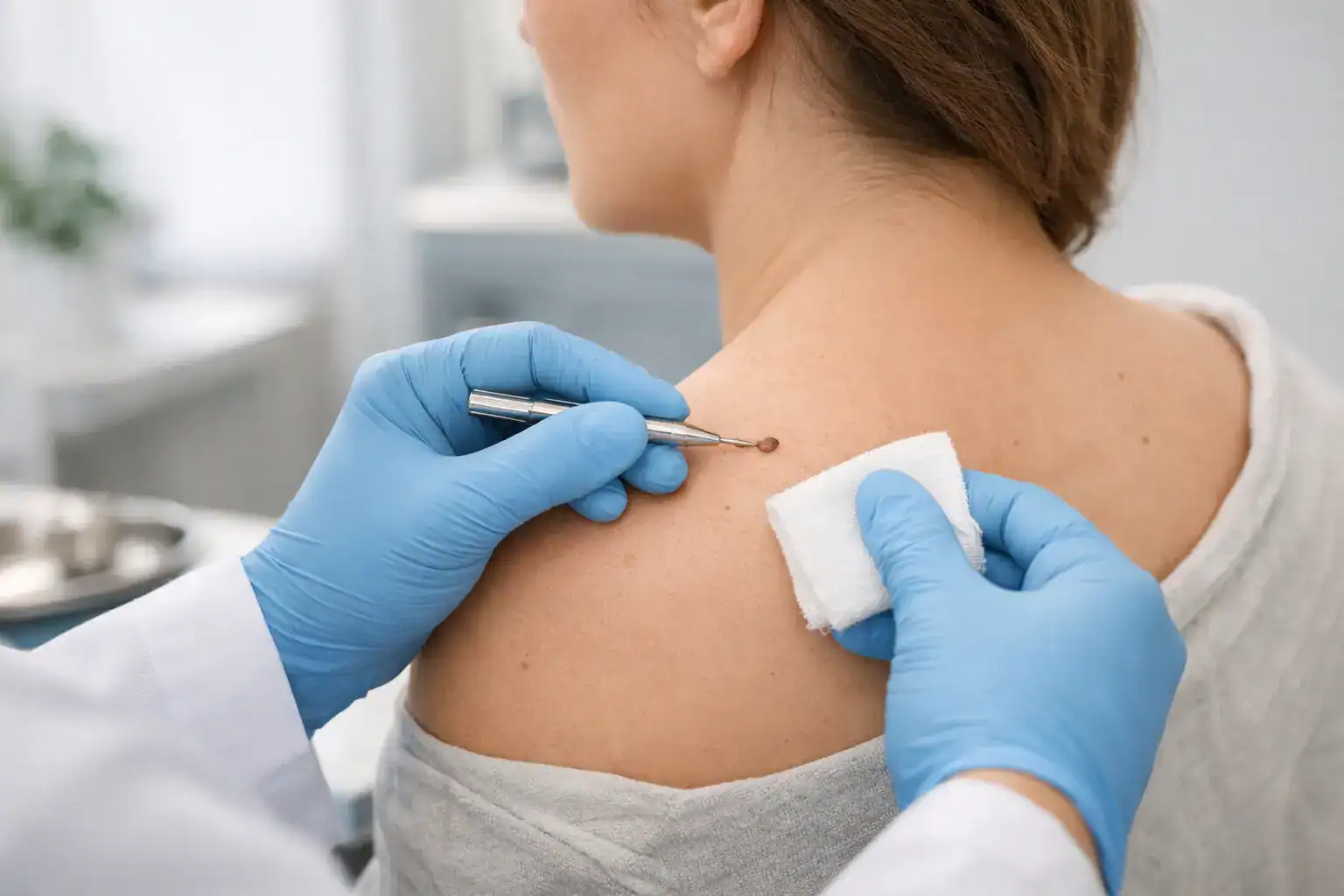
What is a mole biopsy procedure?
A mole biopsy procedure is a minor in-office procedure used to remove all or part of a mole so it can be examined under a microscope. Dermatologists use biopsies to evaluate moles that appear atypical, irritated, changing, or otherwise suspicious for skin cancer, including melanoma.
Not every unusual mole is cancerous. In fact, many biopsied moles turn out to be benign or mildly atypical. Still, visual examination alone has limits. A biopsy provides the tissue diagnosis needed to confirm whether a spot is harmless, precancerous, or cancerous.
That distinction matters because the next step depends on the pathology report. Some moles require no additional treatment after biopsy. Others may need a wider excision, closer monitoring, or a more specialized skin cancer treatment plan.
When a dermatologist may recommend a mole biopsy
Dermatologists do not biopsy every mole, and that is by design. The decision is based on the clinical exam, the mole’s history, and whether it shows warning signs. Common reasons for biopsy include asymmetry, irregular borders, uneven color, increasing diameter, or recent evolution. A mole may also be sampled if it bleeds, itches, becomes tender, or looks clearly different from a patient’s other spots.
A biopsy may also be appropriate when a patient has significant risk factors, such as a personal or family history of melanoma, many atypical moles, or heavy cumulative sun exposure. In those situations, a dermatologist may have a lower threshold to test a mole that seems borderline.
Sometimes the concern is skin cancer. Sometimes it is chronic irritation from clothing, shaving, or friction. Either way, the purpose is to stop guessing and get an answer based on pathology, not appearance alone.
Types of mole biopsy procedure techniques
There is no single biopsy method that fits every mole. The right approach depends on the mole’s size, depth, location, and how suspicious it appears.
Shave biopsy
In a shave biopsy, the dermatologist removes the raised or superficial portion of the mole using a sterile blade after numbing the area. This method is often used for lesions that appear confined to the top layers of skin.
Shave biopsies can be efficient and heal well, especially on the trunk or extremities. The trade-off is that they may not capture the full depth of a deeper lesion, so they are not always the first choice when melanoma is a concern.
Punch biopsy
A punch biopsy uses a circular instrument to remove a small, deeper core of skin. This technique samples both the surface and deeper layers and may be useful for smaller lesions or when depth matters.
Depending on the size, the site may be closed with a stitch or two. Punch biopsies can leave a small round scar, but they often provide more complete tissue architecture for diagnosis.
Excisional biopsy
An excisional biopsy removes the entire mole, usually with a narrow margin of surrounding skin. When a mole is highly suspicious for melanoma, this is often the preferred approach because it gives the pathologist the most complete specimen.
This method typically requires sutures and may leave a longer linear scar than a shave or punch biopsy. Even so, complete removal at the time of biopsy can be the most appropriate medical choice when diagnostic accuracy is the priority.
What happens during the appointment
Most mole biopsies are done in a standard office visit and take only a short time once the procedure begins. After the skin is cleaned, the dermatologist injects a local anesthetic to numb the area. Patients usually feel a quick sting or burn with the numbing medicine, followed by pressure rather than sharp pain during the biopsy itself.
Once the area is numb, the dermatologist removes part or all of the mole using the selected technique. If needed, the wound is closed with sutures. The site is then dressed, and patients receive aftercare instructions before leaving.
The visit is generally efficient, but it should not feel rushed. A good biopsy appointment includes time to explain why the lesion is being tested, what method is being used, and what kind of scar or recovery to expect.
Does a mole biopsy hurt?
Most patients tolerate the procedure very well. The injection of local anesthetic is usually the most noticeable part. After that, discomfort is typically minimal.
Once the numbness wears off, the area may feel sore, tight, or mildly tender for a day or two. Larger biopsies or biopsies in areas with movement, such as the back, chest, or legs, may be more noticeable than small biopsies on less mobile skin. Over-the-counter pain relief is often enough if any discomfort develops.
Pain that steadily worsens, significant swelling, spreading redness, or drainage is not typical and should prompt a call to the dermatology office.
Preparing for a mole biopsy procedure
Preparation is usually simple. Patients should arrive with clean skin and be ready to discuss medications, allergies, and any history of bleeding problems or prior skin cancers. If the lesion is in a hair-bearing area, there is no need to shave it at home unless the office specifically advises that.
Blood thinners do not always need to be stopped, and patients should not make medication changes on their own. The right decision depends on the reason the medication is being taken and the size of the planned biopsy. Your dermatologist can coordinate with other physicians if needed.
It also helps to ask practical questions before the procedure. Will stitches be placed? Are there activity restrictions? Could the biopsy affect work, exercise, or sports for a few days? For busy families and working adults, those details can matter as much as the diagnosis itself.
Recovery and wound care
Healing depends on the biopsy type, the size of the specimen, and the location on the body. A shallow shave biopsy may heal within a couple of weeks, while a deeper excision with sutures may take longer and require a follow-up visit for stitch removal.
Most patients are advised to keep the site clean, apply a thin layer of ointment if directed, and cover it with a fresh bandage until it has sealed over appropriately. Good wound care supports healing and may improve the final scar.
Scarring is possible with any biopsy. That does not mean the scar will be severe, but it is important to be realistic. On the face, chest, shoulders, and back, scar behavior can be less predictable. Skin tone, personal healing tendencies, and biopsy depth all play a role.
When results come back
After the sample is sent to pathology, the tissue is examined under a microscope by a dermatopathologist or pathologist. Results often return within several days to two weeks, though timing can vary.
The report may describe the mole as benign, atypical, dysplastic, precancerous, or malignant. If melanoma or another skin cancer is found, the next step depends on the diagnosis, the margins, and the depth of the lesion. Some cases require only complete excision. Others may call for more involved treatment and close follow-up.
This is one reason specialist access matters. In a practice with medical, surgical, and skin cancer expertise under one system, patients can move from diagnosis to treatment planning without unnecessary delays.
When to be concerned after a biopsy
A small amount of redness or spotting can be normal early on. What is less normal is heavy bleeding that does not stop with pressure, increasing warmth, pus-like drainage, or fever. Those changes can suggest infection or another issue that needs prompt evaluation.
Patients should also follow up if a site does not seem to heal, if a pigmented spot appears to return, or if pathology recommendations are unclear. Clear communication after biopsy is part of good dermatologic care, not an extra.
Why timely evaluation matters
Many suspicious moles turn out to be noncancerous, and that is reassuring. But when a lesion is melanoma or another form of skin cancer, early diagnosis can make treatment simpler and outcomes better. Waiting months to recheck a changing mole is rarely the best plan.
For patients across North Georgia balancing work, family, and health needs, convenience matters too. Access to a nearby dermatology office, timely scheduling, and coordinated follow-up can make it easier to act on a changing mole instead of putting it off.
If a dermatologist recommends a biopsy, it does not automatically mean the spot is dangerous. It means the next smart step is getting a precise answer so your care can be based on evidence, not uncertainty. That clarity is often what gives patients the most peace of mind.