A spot that bleeds when you wash your face, a sore that never quite heals, or a shiny bump that seems harmless can all raise the same question: could this be skin cancer? This basal cell carcinoma guide is designed to help patients understand what basal cell carcinoma is, what it looks like, how it is treated, and when it makes sense to schedule a dermatology visit.
Basal cell carcinoma, often called BCC, is the most common type of skin cancer. It begins in the basal cells, which are found in the lower part of the epidermis. In many cases, it grows slowly and is highly treatable, especially when found early. Even so, it should not be ignored. Left untreated, basal cell carcinoma can continue to enlarge, damage nearby tissue, and become more complicated to remove.
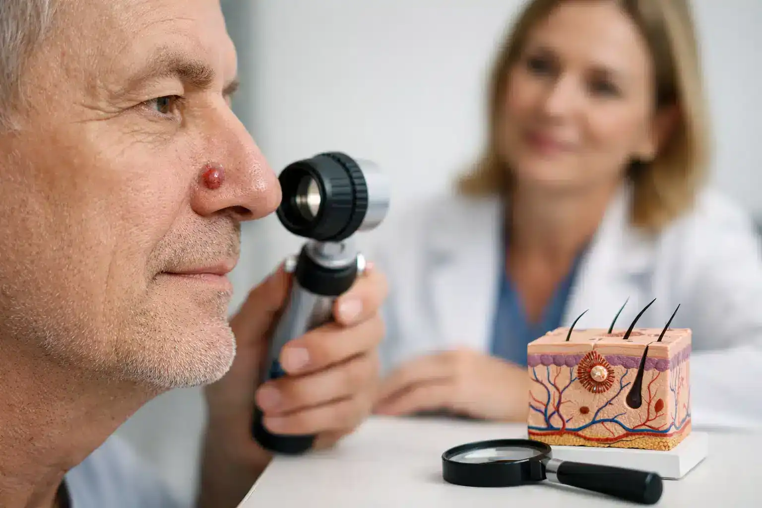
What basal cell carcinoma usually looks like
One reason BCC gets missed is that it does not always look dramatic. Some patients expect skin cancer to appear dark, irregular, or obviously dangerous. Basal cell carcinoma often looks much subtler than that.
It may appear as a pearly or translucent bump, a pink patch, a sore that crusts or bleeds and returns, or a scar-like area that feels firm or waxy. In some skin tones, it can look pink or red. In deeper skin tones, it may appear brown, black, or slightly shiny, which can make it easier to confuse with benign growths or post-inflammatory pigmentation.
Basal cell carcinoma is common on sun-exposed areas such as the face, ears, scalp, neck, chest, shoulders, and arms. That said, it can also develop in places people do not watch closely, especially if they are not doing regular skin checks.
Basal cell carcinoma guide to risk factors
The biggest risk factor is cumulative ultraviolet exposure over time. That includes years of outdoor work, sports, driving, and past sunburns, not just beach vacations. Tanning bed use also increases risk.
Other factors matter too. People with fair skin, light eyes, or a history of frequent sunburns tend to have higher risk, but basal cell carcinoma can occur in any skin tone. Age increases risk because sun damage builds over time, although younger adults can develop BCC as well. A personal history of skin cancer, a weakened immune system, certain genetic syndromes, and prior radiation exposure can all make BCC more likely.
If you have already had one basal cell carcinoma, your chances of developing another are higher. That is why follow-up care and regular skin exams are part of good long-term management, not an extra step.
When to see a dermatologist
If a spot is new, changing, bleeding, scabbing, or not healing after several weeks, it is worth having it examined. The same is true for growths that look different from your other spots or lesions that keep recurring in the same area.
Many patients wait because the lesion is not painful. Pain is not a reliable sign. Basal cell carcinoma can be painless for a long time while still growing beneath the surface.
An in-person evaluation matters because many benign conditions can resemble BCC, and BCC can resemble harmless irritation. A dermatologist can assess the lesion’s pattern, location, and behavior, and determine whether a biopsy is needed.
How diagnosis works
Diagnosis usually begins with a skin exam. If the lesion looks suspicious, the next step is often a biopsy. During a biopsy, the dermatologist removes part or all of the lesion and sends it to a lab for analysis.
This is the only way to confirm whether the spot is basal cell carcinoma. A visual exam can strongly suggest it, but pathology provides the diagnosis and often helps clarify the subtype. That matters because some forms of BCC are more superficial, while others may grow more deeply or have less distinct borders.
For patients, the biopsy process is usually straightforward. The area is numbed, the sample is taken, and aftercare instructions are reviewed. Mild soreness or a small scab is common, but most people return to normal routines quickly.
Treatment options for basal cell carcinoma
The right treatment depends on several factors, including the size of the tumor, its depth, its subtype, its location, whether it has been treated before, and the patient’s overall health. This is where individualized care matters.
Small, superficial basal cell carcinomas may be treated with topical therapy, curettage and electrodesiccation, or other office-based approaches in select cases. These can be effective when the tumor is low-risk and well suited to that method.
Surgical excision is a common treatment and involves removing the tumor along with a margin of surrounding skin. For many BCCs on the trunk or extremities, this is an efficient and reliable option.
Mohs micrographic surgery is often recommended for basal cell carcinomas on cosmetically or functionally important areas such as the nose, eyelids, lips, ears, scalp, and hands, as well as for larger, recurrent, or more aggressive tumors. Mohs allows the surgeon to remove the cancer layer by layer and examine the tissue during the procedure. That approach helps preserve as much healthy tissue as possible while offering very high cure rates.
Some patients may also be candidates for superficial radiotherapy or other specialized treatment strategies when surgery is not the best fit. The trade-offs depend on the clinical picture. A treatment that is appropriate for one patient may not be ideal for another, even if both have BCC.
Why early treatment makes such a difference
Basal cell carcinoma rarely spreads to distant organs, which is reassuring. But that does not mean it is minor. As it grows, it can extend into surrounding skin and deeper tissue. On the face especially, delayed treatment can mean a larger procedure, a more visible scar, and more reconstruction.
Early diagnosis often gives patients more options and may allow for a simpler treatment plan. It can also reduce the chance that the lesion has had time to become recurrent or infiltrative, which can make management more complex.
What recovery and follow-up usually involve
Recovery depends on the treatment used. A biopsy site or small excision may heal with basic wound care and activity modifications for a short period. Mohs surgery can involve more detailed aftercare, especially if stitches or reconstruction are needed.
Patients are usually advised to keep the area clean, follow dressing instructions carefully, and watch for increasing redness, drainage, or pain. Most healing progresses normally, but close communication with your dermatology team helps if questions come up.
Follow-up is just as important as treatment. Once you have had one skin cancer, regular skin exams become part of prevention. Your dermatologist may recommend a schedule based on your history, risk factors, and the number of prior skin cancers.
Preventing future basal cell carcinomas
No prevention plan can reduce risk to zero, but consistent sun protection makes a meaningful difference. Daily broad-spectrum sunscreen, sun-protective clothing, hats, and seeking shade during peak UV hours all help limit additional damage. Avoiding tanning beds is essential.
Patients often ask whether sunscreen still matters after years of sun exposure. The answer is yes. Sun damage is cumulative, and reducing ongoing exposure still helps protect your skin.
Home skin checks are also useful. Look for spots that are changing, bleeding, crusting, or refusing to heal. If something looks off, it is better to have it checked than to wait for certainty.
Choosing the right care team
A good basal cell carcinoma guide should do more than describe the condition. It should help patients understand what to do next. In practical terms, that means choosing a dermatology team with experience in skin cancer diagnosis, biopsy, surgical treatment, and long-term surveillance.
For patients in North Georgia, access and coordination matter too. Skin cancer care is easier to manage when screening, pathology review, surgery, and follow-up are all part of a connected dermatology system. Goodman Dermatology provides comprehensive skin cancer care, including full-body skin exams, in-office procedures, and advanced surgical treatment options designed around both medical outcomes and patient convenience.
If you have a spot that is changing or simply not healing, trust that instinct and get it evaluated. The most helpful next step is often the simplest one: a timely skin exam with an experienced dermatologist.