A mole that looked ordinary last summer can look different by fall. That change is often what gets people to finally book a skin check, and for good reason. When melanoma is found early, treatment is typically simpler and outcomes are often better.
People often search for melanoma early signs photos because they want a quick visual answer. Photos can help, but they have limits. Melanoma does not always look dramatic, and it does not always follow a textbook pattern. A spot can appear small, subtle, or easy to dismiss, especially in its earliest stage.
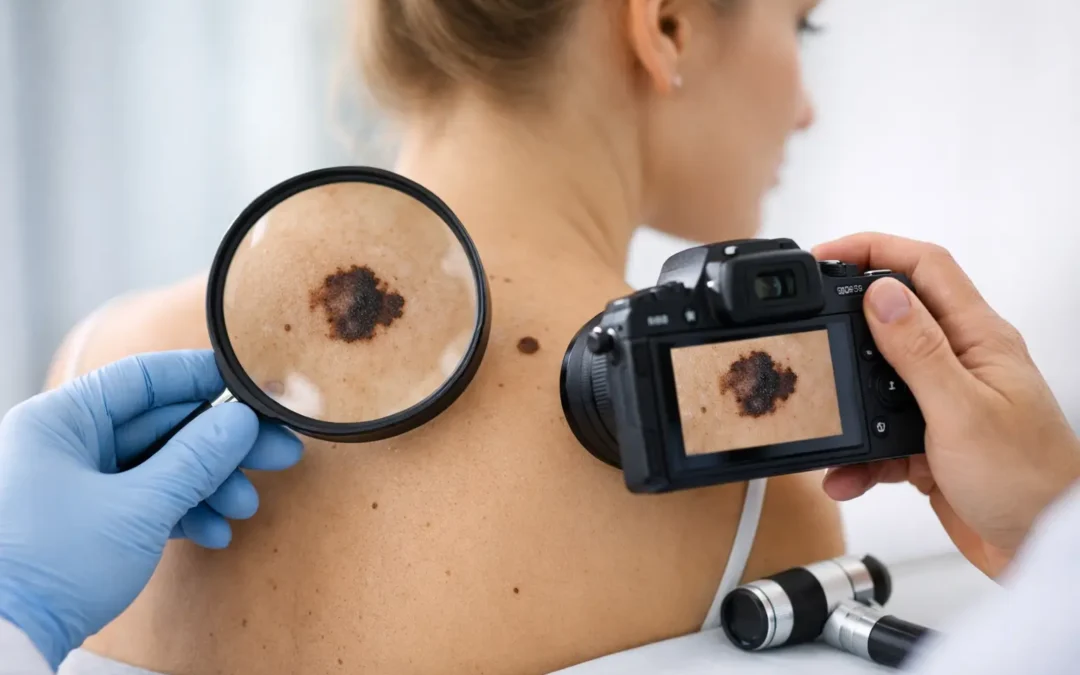
This is where a trained skin cancer evaluation matters. Pictures can raise your awareness. They should not replace an exam by a dermatology professional, especially if a lesion is changing, bleeding, or simply looks different from your other moles.
What melanoma early signs photos can show
Photos are useful because they teach pattern recognition. When you look at examples of early melanoma, you start to notice certain warning signs that deserve attention.
One of the best known tools is the ABCDE rule. In photos, melanoma may appear asymmetrical, meaning one half does not match the other. Borders may look irregular, blurred, jagged, or uneven. Color can vary within the same spot, with shades of brown, black, tan, red, white, or even blue-gray. Diameter can matter, but smaller melanomas do occur, so size alone is not enough. Evolution, or change over time, is often the most important sign.
That last point is worth slowing down for. Many harmless moles stay stable for years. A spot that starts growing, darkening, itching, crusting, or becoming raised deserves prompt attention, even if it does not look alarming in a photo.
Why photos are helpful but not definitive
Images online can create a false sense of certainty in both directions. Some people see a suspicious lesion, compare it with photos, and assume it is probably fine because it does not match exactly. Others become alarmed by a benign mole that happens to resemble one melanoma image.
Both reactions are understandable. The challenge is that melanoma has more than one visual presentation. Some lesions are dark and irregular. Others are pink, red, or nearly skin-colored. Some are flat. Others are raised. Some appear in places people check often, like the arms or face. Others show up on the back, scalp, nails, palms, soles, or between the toes.
Lighting, camera quality, skin tone, and image editing can also affect what a spot looks like in a photo. That is one reason dermatologists rely on more than appearance alone. A full-body skin exam, history of change, and dermoscopic evaluation give a much clearer picture.
The early melanoma signs people miss most often
The most commonly missed sign is change. A mole that becomes darker over a few months, a freckle that develops an irregular edge, or a new spot that keeps enlarging should not be ignored.
Another often-missed sign is the “ugly duckling” mole. This is a lesion that does not look like your other moles. If most of your spots are small, round, and evenly pigmented, but one looks larger or more irregular, that outlier deserves attention.
People also overlook new spots in adulthood. While adults can certainly develop harmless growths, a new pigmented lesion should be watched carefully, especially if it changes quickly. Bleeding without a clear injury, persistent tenderness, or a sore that does not heal are also red flags.
Not all melanomas are dark brown or black
This is where photos can be misleading. Many people expect melanoma to look very dark, but some melanomas are amelanotic, meaning they have little to no pigment. They may appear pink, red, flesh-toned, or slightly inflamed. Because they do not fit the classic image of a dangerous mole, diagnosis can be delayed.
If a pink spot keeps growing, becomes crusted, or does not heal, it still needs evaluation. A lesion does not have to be dark to be serious.
Location matters more than many people realize
Melanoma can occur anywhere on the skin. In men, the back is a common site. In women, the legs are frequently affected. But that pattern is not a rule. Melanoma can also develop on the scalp, ears, under the nails, and on the palms or soles.
These less visible areas are exactly why self-checks and routine professional skin exams work well together. You may catch obvious changes at home, while a dermatologist can examine harder-to-see areas thoroughly.
How to use melanoma early signs photos the right way
Think of photos as a screening prompt, not a diagnosis tool. They are most useful when you compare them with your own skin over time, not when you try to match every detail exactly.
If you are checking your skin at home, good lighting helps. A mirror and a second person can help with the back, scalp, and other difficult areas. Taking a clear photo of a spot can also be helpful if you are monitoring for change, but it should not delay an appointment if the lesion already looks suspicious.
Try to notice three things: whether a spot is new, whether it looks different from your other moles, and whether it is evolving. That approach is often more practical than focusing only on diameter or color.
When a mole needs a dermatologist, not another photo search
If a lesion is changing, bleeding, itching persistently, or looks distinctly different from nearby moles, it is time for a professional evaluation. The same is true if a spot appears after age 30 and continues to evolve, or if you have a personal or family history of skin cancer.
People at higher risk should be especially cautious. That includes those with fair skin, a history of blistering sunburns, frequent tanning bed use, many moles, atypical moles, or immune suppression. But melanoma can affect people of all skin tones, and darker skin does not eliminate risk. In skin of color, melanomas may be more likely to appear on the hands, feet, or under the nails, where they can be missed.
A biopsy is the only way to confirm whether a lesion is melanoma. That can sound intimidating, but in many cases it is a quick in-office procedure. If melanoma is diagnosed, next steps depend on the depth and type of lesion. Early-stage melanoma is often treated effectively when addressed without delay.
What to expect at a skin cancer evaluation
For many patients, the hardest part is simply making the appointment. The exam itself is straightforward. A dermatology provider will review your history, ask whether the lesion has changed, and examine the area closely. A full-body skin exam may be recommended, especially if you have multiple moles or significant sun exposure history.
If a spot appears suspicious, your provider may recommend a biopsy. That step is not taken lightly, but it is the most reliable way to get an answer. Waiting and watching can be reasonable for some benign-appearing lesions, but if melanoma is part of the concern, early diagnosis matters.
At Goodman Dermatology, patients across North Georgia have access to expert skin cancer evaluation, full-body skin exams, and advanced treatment options when needed. That combination of specialist care and convenient local access can make it easier to act quickly when a spot does not look right.
A practical approach for patients and families
For busy adults, parents, and older patients, the simplest system is often the best one. Check your skin on a regular basis, pay attention to new or changing lesions, and do not rely on online images alone to reassure you.
Photos can be educational. They can even be the reason someone catches melanoma early. But there is always a trade-off with self-assessment. The more you search, the easier it is to either normalize something suspicious or worry about something harmless. A timely dermatology visit removes that guesswork.
If you see a mole or spot that is evolving, looks irregular, or stands out from the rest, trust that observation. Early melanoma is not always dramatic, and that is exactly why prompt evaluation matters.
The most helpful next step is often the simplest one: if your skin is showing you something new or changing, let a dermatology expert take a closer look.